
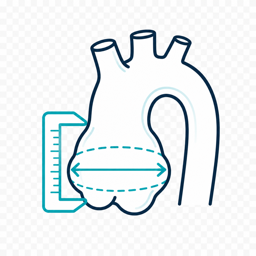

Imaging surveillance
Establish a baseline cardiovascular assessment and use serial echocardiography when appropriate. Consider more frequent imaging when dilation, rapid growth, or other high-risk features are present.
Use CMR or CT when TTE is inadequate or when assessment of the distal aorta is needed.